a b Fig. 3.50. A child with acute onset of respiratory distress.(See Appendix 2) a b Fig. 3.51. Chronic lung disease.(See Appendix 2) way to read systematically is to evaluate the tech- and diaphragm). Now evaluate Figs.3.50-3.52.- nical factors of lung volume, patient position, and proach each one systematically and describe the - the way the ?lm was exposed. Use the radiologist's normalities you see. The answers are in the App- circle and ABCS: A=abdomen, B=bones, S=soft dix 2. tissue, and C=chest (airway, mediastinum, lungs, 03_Haller_Chest 22.10.2004 13:06 Uhr Seite 62 62 3 Chest Examinations in Children a b c d Fig. 3.52. A child with wheezing.(See Appendix 2) 3. Webb WR, Mueller NL, Naidich DP (1996) High-resolu- References and Further Reading tion CT of the lung,2nd edn.Lippincott-Raven,New York 4. Kuhn JP,Slovis TL,Haller JO (eds) (2003) Caffey's pedi- 1. Slovis TL, Haller JO, Berdon WE, Baker DH, Joseph PM atric diagnostic imaging,10th edn.Mosby,Philadelphia (1979) Noninvasive visualization of the pediatric airway.
Dieser Download kann aus rechtlichen Gründen nur mit Rechnungsadresse in A, B, BG, CY, CZ, D, DK, EW, E, FIN, F, GR, HR, H, IRL, I, LT, L, LR, M, NL, PL, P, R, S, SLO, SK ausgeliefert werden.
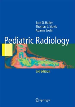
Aus den Rezensionen zur 3. Auflage: " Jedes Kapitel enthält eine fülle von Bildbeispielen, die dem Unerfahrenen die Pathologien und Besonderheiten demonstrieren und das Wissen des Erfahrenen bereichern. Eine besondere Qualität des Buches stellen die Quizfragen dar, die anhand von Bildmaterial und klinischen Beschreibungen die Kenntnisse des Lesers testen. Darüber hinaus enthält es wertvolle praktische Hinweise zur Planung und Interpretation pädiatrischer Bildgebung. Differenzialdiagnostische Tabellen und klare anatomische Zeichnungen unterstützen die Bildinterpretation. Es stellt eine exzellente Ausbildungsmöglichkeit in der pädiatrischen Radiologie dar." (in: Paediatrica, 2007, Vol. 18, Issue 4, S. 70)

