140,99 €
140,99 €
inkl. MwSt.
Sofort per Download lieferbar
payback
0 °P sammeln
140,99 €
140,99 €
inkl. MwSt.
Sofort per Download lieferbar

Alle Infos zum eBook verschenken
payback
0 °P sammeln
Als Download kaufen
140,99 €
inkl. MwSt.
Sofort per Download lieferbar
payback
0 °P sammeln
Jetzt verschenken
140,99 €
inkl. MwSt.
Sofort per Download lieferbar

Alle Infos zum eBook verschenken
payback
0 °P sammeln
  • Format: ePub

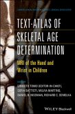
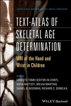
The first complete textbook and atlas of the vitally important technique of bone age assessment utilizing MRI for children's hand and wrist This latest volume in the growing Wiley Current Clinical Imaging series is a must-have resource that collects, in a single volume, all that is currently known and applicable about the use of magnetic resonance imaging (MRI) for the assessment of bone age. Presented in two parts, Text-Atlas of Skeletal Age Determination: MRI of the Hand and Wrist in Children first focuses on the anatomic, social, and legal aspects of bone age, providing a concise overview…mehr

  • Geräte: eReader
  • mit Kopierschutz
  • eBook Hilfe
  • Größe: 16.92MB
Produktbeschreibung
The first complete textbook and atlas of the vitally important technique of bone age assessment utilizing MRI for children's hand and wrist This latest volume in the growing Wiley Current Clinical Imaging series is a must-have resource that collects, in a single volume, all that is currently known and applicable about the use of magnetic resonance imaging (MRI) for the assessment of bone age. Presented in two parts, Text-Atlas of Skeletal Age Determination: MRI of the Hand and Wrist in Children first focuses on the anatomic, social, and legal aspects of bone age, providing a concise overview of the use of bone age determination in medical, legal, and social systems.??It then covers the clinical use and application of MRI in assessing bone age. The book offers complete chapter coverage on endocrinology, puberty, and disorders of pubertal development; bone marrow maturation in healthy and diseased states; growth failure and pediatric inflammatory bowel disease; skeletal findings in neurometabolic disease, genetic disease, and pediatric oncology patients; and much more. Text-Atlas of Skeletal Age Determination provides: * A comprehensive review of the medical, legal, and social aspects of bone age assessment * An in-depth discussion of MRI as an alternative to the traditional ionizing radiation-based radiographic techniques for the assessment of bone age * Complete guidelines for clinical application of these MRI-based techniques * "Recipes" for replicating these techniques and applications for diverse patient populations * Cutting-edge information prepared and presented by an international team of experts * A superb collection of beautifully reproduced, high-quality images This is an ideal book for radiologists, pediatricians, family physicians, endocrinologists, and sports medicine physicians interested in skeletal development and bone age assessment.

Dieser Download kann aus rechtlichen Gründen nur mit Rechnungsadresse in A, B, BG, CY, CZ, D, DK, EW, E, FIN, F, GR, HR, H, IRL, I, LT, L, LR, M, NL, PL, P, R, S, SLO, SK ausgeliefert werden.

Autorenporträt
Ernesto Tomei, MD, is Professor of Radiology in the Department of Radiological Sciences, Oncology, and Pathological Anatomy at the "Sapienza" University of Rome in Rome, Italy. He was a visiting lecturer in 1988-89 in the Department of Radiology at the University of California San Francisco (USA), as well as in the Department of Radiology at the University of Berlin (Klinikum Charlottenburg). He is an international leader in the emerging use of MRI for bone age assessment, and is currently engaged in a major project on this topic, which is co-financed by the European Union and the Italian Ministry of the Interior. He is also heavily involved in teaching residents specializing in Internal Medicine and Radiology and is an associate editor or reviewer for several European radiology and internal medicine journals. Richard C. Semelka, MD, is one of the foremost authorities on MRI of the abdomen and pelvis, with six MRI text-books and over 200 peer-reviewed papers to his credit. Dr. Semelka has also authored numerous chapters and review articles on MRI of the body, and has presented his work at meetings around the world. Dr. Semelka is the Director of MR Services, Professor and Vice Chair for Research, Department of Radiology, University of North Carolina at Chapel Hill. Daniel Nissman, MD, is an Assistant Professor of Radiology at the University of North Carolina at Chapel Hill, USA. He is primarily a musculoskeletal radiologist with experience in bone age assessment using both ionizing and non-ionizing radiologic modalities.