48,95 €
48,95 €
inkl. MwSt.
Sofort per Download lieferbar
payback
24 °P sammeln
48,95 €
48,95 €
inkl. MwSt.
Sofort per Download lieferbar

Alle Infos zum eBook verschenken
payback
24 °P sammeln
Als Download kaufen
48,95 €
inkl. MwSt.
Sofort per Download lieferbar
payback
24 °P sammeln
Jetzt verschenken
48,95 €
inkl. MwSt.
Sofort per Download lieferbar

Alle Infos zum eBook verschenken
payback
24 °P sammeln
  • Format: ePub

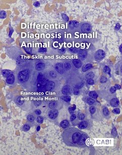
Illustrated with high-quality photomicrographs, Differential Diagnosis in Small Animal Cytology: The Skin and Subcutis is a comprehensive resource for identifying through cytology the most common cutaneous and subcutaneous diseases of dogs and cats. With key points describing the main clinical and cytological features of each lesion, the book provides lists of differential diagnoses, including diagnostic algorithms, and handy 'hints and tips' boxes. It is also enriched by chapters on the correct use and maintenance of the microscope, and techniques of collection and preparation of cytological…mehr

  • Geräte: eReader
  • mit Kopierschutz
  • eBook Hilfe
  • Größe: 20.5MB
Produktbeschreibung
Illustrated with high-quality photomicrographs, Differential Diagnosis in Small Animal Cytology: The Skin and Subcutis is a comprehensive resource for identifying through cytology the most common cutaneous and subcutaneous diseases of dogs and cats. With key points describing the main clinical and cytological features of each lesion, the book provides lists of differential diagnoses, including diagnostic algorithms, and handy 'hints and tips' boxes. It is also enriched by chapters on the correct use and maintenance of the microscope, and techniques of collection and preparation of cytological specimens, making the book a valuable resource for veterinary pathologists (clinical and anatomic), residents, veterinary undergraduate students and small animal practitioners. Key features: -Over 130 photomicrographs of the most common skin and subcutaneous lesions to help with diagnosis. -Ideal reference book with concise descriptions of each lesion. -Organised into key bullet points to facilitate use during diagnostic work, or as a revision aid.

Dieser Download kann aus rechtlichen Gründen nur mit Rechnungsadresse in A, D ausgeliefert werden.

Autorenporträt
Francesco Cian qualified from University of Padua (Italy) with a DVM in 2006 and spent the next 4 years in small animal practice. In 2010, he started a residency program in Clinical Pathology at the University of Cambridge, which he finished in 2013 attaining both an ECVCP and FRCPath diploma. Francesco joined the Animal Health Trust (AHT) in 2013 as Head of Clinical Pathology, and from September 2015 works for Battlab (LABOKLIN). Francesco has a special interest in lymphoproliferative disorders of dogs and cats and flow cytometry. He is member of the cytology exam committee of the European College of Veterinary Clinical Pathology (ECVCP) and is author of several publications on peer-reviewed journals. He co-authored with Paola Monti the cytology chapter of the third edition of the BSAVA Manual of Veterinary Clinical Pathology and is editor of the second edition of the book Veterinary Cytology: Dog, Cat, Horse and Cow: Self-Assessment Color Review.