142,95 €
142,95 €
inkl. MwSt.
Sofort per Download lieferbar
payback
71 °P sammeln
142,95 €
142,95 €
inkl. MwSt.
Sofort per Download lieferbar

Alle Infos zum eBook verschenken
payback
71 °P sammeln
Als Download kaufen
142,95 €
inkl. MwSt.
Sofort per Download lieferbar
payback
71 °P sammeln
Jetzt verschenken
142,95 €
inkl. MwSt.
Sofort per Download lieferbar

Alle Infos zum eBook verschenken
payback
71 °P sammeln
  • Format: ePub

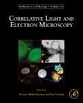
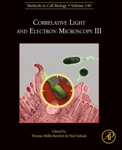
Correlative Light and Electron Microscopy III, Volume 140 , a new volume in the Methods in Cell Biology series, continues the legacy of this premier serial with quality chapters authored by leaders in the field. Topics discussed in this new release include Millisecond time-resolved CLEM, Super resolution LM und SEM of high-pressure frozen C. elegans, Preservation fluorescence, super res CLEM, APEX in Tissue, Corrsight mit IBIDI flowthrough chamber, Correlative Light Atomic Force Electronic Microscopy (CLAFEM), Atmospheric EM CLEM, and High-precision correlation, amongst other…mehr

  • Geräte: eReader
  • mit Kopierschutz
  • eBook Hilfe
  • Größe: 189.98MB
Produktbeschreibung
Correlative Light and Electron Microscopy III, Volume 140, a new volume in the Methods in Cell Biology series, continues the legacy of this premier serial with quality chapters authored by leaders in the field. Topics discussed in this new release include Millisecond time-resolved CLEM, Super resolution LM und SEM of high-pressure frozen C. elegans, Preservation fluorescence, super res CLEM, APEX in Tissue, Corrsight mit IBIDI flowthrough chamber, Correlative Light Atomic Force Electronic Microscopy (CLAFEM), Atmospheric EM CLEM, and High-precision correlation, amongst other topics.

Chapters in this ongoing series deal with different approaches for analyzing the same specimen using more than one imaging technique. The strengths and application area of each is presented, with this volume exploring the aspects of sample preparation of diverse biological systems for different CLEM approaches.

  • Contains contributions from experts in the field
  • Covered topics include targeted ultramicrotomy and high-precision correlation
  • Presents recent advances and currently applied correlative approaches
  • Gives detailed protocols allowing the application of workflows in one's own laboratory setting
  • Covers CLEM approaches in the context of specific applications
  • Aims to stimulate the use of new combinations of imaging modalities

Dieser Download kann aus rechtlichen Gründen nur mit Rechnungsadresse in A, B, BG, CY, CZ, D, DK, EW, E, FIN, F, GR, HR, H, IRL, I, LT, L, LR, M, NL, PL, P, R, S, SLO, SK ausgeliefert werden.

Autorenporträt
Thomas Müller-Reichert is a Professor of Structural Cell Biology at the Technische Universität Dresden (TU Dresden, Germany). He is interested in how the microtubule cytoskeleton is modulated within cells to fulfill functions in mitosis, meiosis and abscission. The Müller-Reichert lab is mainly applying correlative light microscopy and electron tomography to study the 3D organization of microtubules in early embryos and meiocytes of the nematode Caenorhabditis elegans, and also in mammalian cells in culture. He has published over 75 papers and edited several volumes of the Methods in Cell Biology series on electron microscopy and CLEM.

TMR obtained his PhD at the Swiss Federal Institute of Technology (ETH) in Zurich and moved afterwards for a post-doc to the EMBL in Heidelberg (Germany). He was a visiting scientist with Dr. Kent McDonald (UC Berkeley, USA). Together with Paul Verkade, he set up the electron microscope facility at the newly founded Max Planck Institute of Mo

lecular Cell Biology and Genetics (MPI-CBG). Since 2010 he is a scientific group leader and head of the Core Facility Cellular Imaging (CFCI) of the Faculty of Medicine Carl Gustav Carus of the TU Dresden. He acted as president of the German Society for Electron Microscopy (Deutsche Gesellschaft für Elektronenmikroskopie, DGE) from 2018 to 2019.
He taught numerous courses and workshops on high-pressure freezing and Correlative Light and Electron Microscopy.