161,99 €
inkl. MwSt.
Versandkostenfrei*
Versandfertig in über 4 Wochen
payback
81 °P sammeln
  • Gebundenes Buch

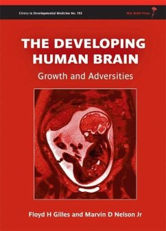
This book is about human brain development, focusing on the last half of gestation and the neonatal and infant periods. These periods bring the greatest risk for the acquisition of childhood functional neurologic deficits, including cerebral palsy, developmental delay and intellectual disability. Section 1 covers typical development, including growth in brain weight, ventricular surfaces, gyral development, myelinated tract development, magnetic resonance spectroscopy, and angiogenesis, all serving as reference points for section 2, which deals with common acquired brain abnormalities, some of…mehr

Produktbeschreibung
This book is about human brain development, focusing on the last half of gestation and the neonatal and infant periods. These periods bring the greatest risk for the acquisition of childhood functional neurologic deficits, including cerebral palsy, developmental delay and intellectual disability. Section 1 covers typical development, including growth in brain weight, ventricular surfaces, gyral development, myelinated tract development, magnetic resonance spectroscopy, and angiogenesis, all serving as reference points for section 2, which deals with common acquired brain abnormalities, some of which are often underemphasized or overlooked. The topics in section 2 include retrocerebellar cysts, abnormal events in fetal brain, white-matter abnormalities, lesions of gray and white matter, hemorrhage, ventriculomegaly and hydrocephalus, late expressions of fetal brain disease, and reactions of the developing brain to chronic disease. Between sections 1 and 2 is a chapter on embryonic
and fetal physiologic reactions to external stimuli. Where appropriate, the authors have combined pathologic with neuroimaging examples to help the reader better understand the neuroimages that they encounter.
Much of the information in the book is based on data from the National Collaborative Perinatal Project, still the only large autopsy survey of late fetal brain lesions
Autorenporträt
Floyd Gilles heads the pediatric neuropathology program at the Children's Hospital Los Angeles. He is the Burton E. Green Professor of Pediatric Neuropathology at CHLA, and Professor of Pathology (Neuropathology), Neurology, and Neurosurgery at the Keck School of Medicine, University of Southern California. His areas of expertise include the intraoperative interpretation of frozen sections of brain specimens and the neuropathologic aspects of childhood autopsies. His major areas of research interest include the neuropathology of the last half of gestation, pediatric brain tumor identification, classification, prognosis and therapeutic trials; the development of a statistically modeled method of classifying tumors that provides specific five-year survival probabilities and appropriate survival covariates; and telencephalic angiogenesis. Marvin Nelson is Chief of the Department of Imaging Services and the John L. Gwinn Professor of Pediatric Radiology at CHLA, and Professor of Radiology at the Keck School of Medicine. He also heads the training program in pediatric radiology and neuroradiology at CHLA. He was a fellow in neuroradiology at the National Hospital for Nervous Diseases, Queen Square, London and the Rothschild Institute, Paris, France, and a fellow in pediatric neuroradiology at Children's Memorial Hospital, Chicago, Illinois. He joined the faculty at CHLA and USC in 1987. His research includes the use of spectroscopy and other advanced imaging techniques in diagnosing such conditions as brain tumors, pediatric acute hydrocephalus and sickle cell disease damage.
Rezensionen
"The text is comprehensive, well referenced and supported by numerous illustrations, including pictures, tables and diagrams. This is a useful resource that takes the reader into a fascinating journey of exploration." (Nursing Standard, 31 October 2012)