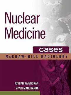
Publisher's Note: Products purchased from Third Party sellers are not guaranteed by the publisher for quality, authenticity, or access to any online entitlements included with the product. A unique case-based approach to understanding nuclear medicine 176 cases and 1190 illustrations (many in full color) "They have implemented a compelling approach to the case-based format....In summary, you will love this book. It is thoughtfully constructed and reader focused. You will see manifest the inspiration and commitment of the two editors. Enjoy, learn and ultimately have an impact."-Norman J. Beauchamp, University of Washington (from the foreword) Nuclear Medicine Cases features 176 nuclear medicine and PET/CT cases grouped according to organ system. Each case includes presentation, findings, differential diagnosis, comments, pearls, and numerous images, many in full color. Covering a wide range of general clinical topics of interest to practicing imaging physicians, this well-illustrated reference guide covers endocrine, musculoskeletal, chest, genitourinary, gastrointestinal, lymphatic, CNS, renal, vascular cases and includes a separate section for pediatrics. The book's easy-to-navigate organization is specifically designed for use at the workstation. The concise quick-scan text, numerous images, and helpful icons and pearls speed and simplify the learning process. FEATURES: * 176 cases and 1190 illustrations (many in full color) * An icon-indicated grading system depicting the full spectrum of findings from common to rare and typical to unusual, and the consistent chapter organization make this the perfect workstation reference * Emphasizes the latest diagnostic modalities * Covers a wide range of clinical topics About the McGraw-Hill Radiology SeriesThis innovative series offers indispensable workstation reference material for the practicing radiologist. Within this series is a full range of practical, clinically relevant works divided into three categories: . Patterns books: organized by modality, these books provide a pattern-based approach to constructing practical differential diagnosis. Variants books: structured by modality as well as anatomy, these graphic references aid the radiologist in reducing false-positive rates. Cases books: classic case presentations with an emphasis on differential diagnoses and clinical context


