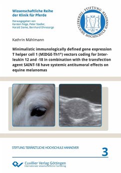
Equine melanomas are the most common neoplasms in older grey horses. Until now, no curative therapy is established. Xenogenic DNA vaccination represents a promising therapeutic approach. Goal of the present study was to evaluate the safety and clinical efficacy of vaccination of grey horses with natural occurring melanomas with MIDGE-Th1® vectors encoding for equine (eq) IL12 and eqILRAP-IL18 alone or in combination with human gp100 (hgp100) or human tyrosinase (htyr). Three groups (each n=9) of adult grey horses with variable numbers of melanomas received three intramuscular and three intradermal peritumoral vaccinations. Each group was treated with MIDGE-Th1® vectors coding for eqIL12 and eqILRAP-IL18. One group (group gp100) was additionally treated with 500 ¿g hgp100MIDGE-Th1® and one group with 500 ¿g htyrMIDGE-Th1® (group tyr). The third group did not receive additional DNA (group IL12/18). Vectors were complexed with the transfection agent SAINT-18.The injections were performed on days 1, 22 and 78 intradermally around one selected melanoma (locally treated melanoma) and intramuscularly as a systemic treatment. A general examination of the horses and evaluation of local reactions at the injection site were performed daily during hospitalisation (on the day of each injection and 3 days after each injection as well as on day 120). In each horse the locally treated melanoma and up to 8 non-locally treated melanomas were measured before each injection and on day 120 by calliper and ultrasound. The relative volumes were calculated in relation to day 1 (100%). For the evaluation of the generation of specific CD8+ cytotoxic T-cells against hgp100 and htyr, peripheral blood mononuclear cells (PBMCs) isolated before each injection and on day 120, were stimulated in vitro with autologous dermal cells transfected with hgp100 or htyr. Interferon ¿ (IFN ¿) as a response to this specific stimulation was measured by intracellular immunofluorescence staining.

