51,99 €
inkl. MwSt.
Versandkostenfrei*
Versandfertig in 6-10 Tagen
payback
26 °P sammeln
  • Broschiertes Buch

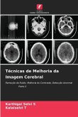
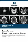
Digital images find their applications in many fields like medical imaging, biometric, multimedia, remote sensing etc. Magnetic Resonance (MR) image is increasingly being used in clinical setting as an adjunct to computed tomography (CT), positron emission tomography (PET) and ultrasound images. MR image provides more spatial information in different contrast and is sensitive to tumor and multifocal disease. Due to its sensitivity, it produces some complications in visual diagnosis that consumes more time and affects therapeutic plans. This book presents a promising set of image analysis…mehr

Produktbeschreibung
Digital images find their applications in many fields like medical imaging, biometric, multimedia, remote sensing etc. Magnetic Resonance (MR) image is increasingly being used in clinical setting as an adjunct to computed tomography (CT), positron emission tomography (PET) and ultrasound images. MR image provides more spatial information in different contrast and is sensitive to tumor and multifocal disease. Due to its sensitivity, it produces some complications in visual diagnosis that consumes more time and affects therapeutic plans. This book presents a promising set of image analysis methods developed for the purpose of assisting radiologists in pre-processing and detecting brain tumor from multimodal image data acquired using MR imaging.
Autorenporträt
O Dr. S. Karthigai Selvi, Faculdade e o Dr. T. Kalaiselvi, Professor Assistente estão a trabalhar no Instituto Rural Gadhigram - Deemed to be University. Têm trabalhado no Processamento de Imagem Médica durante os últimos 10 anos. Os seus trabalhos promissores são publicados em revistas de renome.