- 12%
Beschreibung
Produktdetails
Einband
Taschenbuch
Erscheinungsdatum
16.03.2026
Verlag
LAP LAMBERT Academic PublishingSeitenzahl
80
Maße (L/B/H)
22/15/0,5 cm
Gewicht
137 g
Sprache
Englisch
ISBN
978-620-9-70385-0
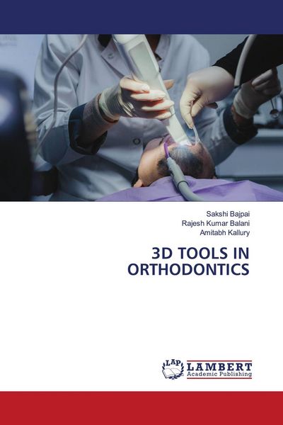
Three-dimensional (3D) imaging tools have brought significant advancements in orthodontic diagnosis, treatment planning, and evaluation of treatment outcomes. Traditionally, orthodontic records were based on two-dimensional (2D) methods such as cephalometric radiographs, panoramic radiographs, clinical photographs, and plaster models. Although these methods have been widely used, they possess inherent limitations, including image distortion, projection errors, inconsistent landmark identification, and difficulty in accurately evaluating complex craniofacial structures. The conversion of three-dimensional anatomical structures into two-dimensional images frequently results in the loss of important diagnostic information and may obscure asymmetries.The introduction of 3D imaging technologies has enabled clinicians to visualize craniofacial structures in their true anatomical dimensions, thereby improving diagnostic accuracy and treatment planning.
Noch keine Bewertungen vorhanden
Verfassen Sie die erste Bewertung zu diesem Artikel
Helfen Sie anderen Kundinnen und Kunden durch Ihre Meinung.
Kurze Frage zu unserer Seite
Vielen Dank für dein Feedback
Wir nutzen dein Feedback, um unsere Produktseiten zu verbessern. Bitte habe Verständnis, dass wir dir keine Rückmeldung geben können. Falls du Kontakt mit uns aufnehmen möchtest, kannst du dich aber gerne an unseren Kund*innenservice wenden.
zum Kundenservice