
KI-Beratung Beta

Gutscheinbedingungen

**Gültig vom 05.10.2026 bis 07.10.2026 | Gültig für nicht preisgebundene fremdsprachige Bücher | Einzelne Artikel können ausgeschlossen sein | Online auf www.bücher.de einlösbar | Click & Collect nur mit Online-Zahlung (Paypal/Kreditkarte) vorab | Nicht kombinierbar mit anderen Gutscheinen oder Preisaktionen | Nur einmal pro Einkauf einlösbar | Gutschein wird auf max. 500€ Bestellwert angerechnet | Keine Barauszahlung | Nicht gültig für Versandkosten und Services

  • Produktbild: Radiology of The Sella Turcica
  • Produktbild: Radiology of The Sella Turcica

Radiology of The Sella Turcica

97,99 €

inkl. gesetzl. MwSt., Versandkostenfrei


Beschreibung

Produktdetails

Einband

Taschenbuch

Erscheinungsdatum

23.11.2011

Abbildungen

XXII, 262 p.

Illustriert von

M. Gaudron

Verlag

Springer Berlin

Seitenzahl

262

Maße (L/B/H)

28/21/1,6 cm

Gewicht

711 g

Auflage

Softcover reprint of the original 1st ed. 1981

Sprache

Englisch

ISBN

978-3-642-67788-5

Beschreibung

Produktdetails

Einband

Taschenbuch

Erscheinungsdatum

23.11.2011

Abbildungen

XXII, 262 p.

Illustriert von

M. Gaudron

Verlag

Springer Berlin

Seitenzahl

262

Maße (L/B/H)

28/21/1,6 cm

Gewicht

711 g

Auflage

Softcover reprint of the original 1st ed. 1981

Sprache

Englisch

ISBN

978-3-642-67788-5

Herstelleradresse

Springer-Verlag GmbH
Europaplatz 3
69115 Heidelberg
DE

Email: ProductSafety@springernature.com

Noch keine Bewertungen vorhanden

Verfassen Sie die erste Bewertung zu diesem Artikel

Helfen Sie anderen Kundinnen und Kunden durch Ihre Meinung.

Kundinnen und Kunden meinen

Bewertungen (0)

  • Produktbild: Radiology of The Sella Turcica
  • Produktbild: Radiology of The Sella Turcica
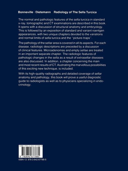
  • 1 Embryology of the Sellar Region.- A. Development of the Sphenoid Bone.- B. Development of the Sphenoid Sinus.- C. Development of the Pituitary Gland.- D. Main Anomalies in the Fetal Development of the Sellar Region.- 2 Anatomy of the Sellar Region.- A. Descriptive Anatomy of the Sellar Region.- B. Relationships Between the Sella Turcica and the Surrounding Structures.- C. Vascular Supply of the Sellar Region.- D. Innervation of the Sellar Region.- 3 Radiographic Techniques.- A. Plain Radiography.- B. Tomography.- 4 Radiologic Anatomy.- A. Radiologic Anatomy of the Sella Turcica and of the Presellar Region.- B. Regional Radiologic Anatomy.- 5 Variations and Normal Limits.- A. Variations in the General Appearance of the Sella Turcica.- B. Variations in Different Anatomic Structures.- 6 Intrasellar Pathology.- A. The Empty Sella Turcica.- B. Pituitary Adenomas.- C. Intrasellar Craniopharyngiomas.- D. Miscellaneous Disorders.- 7 Suprasellar Pathology.- A. Craniopharyngiomas.- B. Hypothalamic Gliomas.- C. Gliomas of the Optic Chiasm.- D. Miscellaneous Disorders.- 8 Presellar Pathology.- A. Gliomas of the Optic Pathways (Nerve and Chiasm).- B. Presellar Meningiomas.- C. Diagnosis of an Abnormal Presellar Region.- 9 Parasellar Pathology.- A. Vascular Disease.- B. Meningiomas.- C. Gasserian Neurinomas and Meningiomas.- D. Diseases of Temporal Lobe.- 10 Retrosellar Pathology.- A. Chordomas.- B. Chondromas.- C. Clivus Meningiomas.- D. Aneurysms of the Basilar Artery.- 11 Infrasellar Pathology.- A. Inflammatory, Infectious, and Mycotic Lesions of the Sphenoid Sinus.- B. Infrasellar Neoplastic Diseases.- 12 Sella Turcica in Raised Intracranial Pressure and Hydrocephalus.- A. Raised Intracranial Pressure.- B. Sella Turcica in Chronic Obstructive Hydrocephalus.- C. Changes in the Sella Turcica in Childhood.- D. Sella Turcica in Craniostenosis.- 13 Generalized Diseases and Changes in the Sella Turcica.- A. Congenital Anomalies of the Sella Turcica.- B. Metabolic Diseases.- C. Endocrine Diseases.- D. Hematologic Diseases.- E. Neoplasms.- F. Infectious Diseases.- G. Fractures of the Sellar Region.- H. Miscellaneous.- 14 Exercises and Pitfalls.- 15 Advances in CT of the Pituitary Gland.- A. Method of CT Examination.- B. Results.- C. Bibliography.- References.