KI-Beratung Beta

  • Produktbild: Methods of Preparation for Electron Microscopy
  • Produktbild: Methods of Preparation for Electron Microscopy

Methods of Preparation for Electron Microscopy An Introduction for the Biomedical Sciences

106,99 €

inkl. gesetzl. MwSt., Versandkostenfrei


Beschreibung

In 1939, when the electron optics laboratory of Siemens & Halske Inc. began to manufacture the first electron microscopes, the biological and medical profes sions had an unexpected instrument at their disposal which exceeded the reso lution of the light microscope by more than a hundredfold. The immediate...

Beschreibung

In 1939, when the electron optics laboratory of Siemens & Halske Inc. began to manufacture the first electron microscopes, the biological and medical profes sions had an unexpected instrument at their disposal which exceeded the reso lution of the light microscope by more than a hundredfold. The immediate and broad application of this new tool was complicated by the overwhelming prob lems inherent in specimen preparation for the investigation of cellular struc tures. The microtechniques applied in light microscopy were no longer appli cable, since even the thinnest paraffin layers could not be penetrated by electrons. Many competent biological and medical research workers expressed their anxiety that objects in high vacuum would be modified due to complete dehydration and the absorbed electron energy would eventually cause degrada tion to rudimentary carbon backbones. It also seemed questionable as to whether it would be possible to prepare thin sections of approximately 0. 5 11m from heterogeneous biological specimens. Thus one was suddenly in posses sion of a completely unique instrument which, when compared with the light microscope, allowed a 10-100-fold higher resolution, yet a suitable preparation methodology was lacking. This sceptical attitude towards the application of electron microscopy in bi ology and medicine was supported simultaneously by the general opinion of colloid chemists, who postulated that in the submicroscopic region of living structures no stable building blocks existed which could be revealed with this apparatus.

Produktdetails

Einband

Taschenbuch

Erscheinungsdatum

12.06.1987

Abbildungen

XVIII, 190 p.

Verlag Springer Berlin

Produktdetails

Einband

Taschenbuch

Erscheinungsdatum

12.06.1987

Abbildungen

XVIII, 190 p.

Verlag Springer Berlin
Seitenzahl

190

Maße (L/B/H)

24,4/17/1,2 cm

Gewicht

375 g

Auflage

Softcover reprint of the original 1st ed. 1987

Übersetzt von

David G. Robinson

Sprache

Englisch

ISBN

978-3-540-17592-6

Herstelleradresse

Springer-Verlag KG
Sachsenplatz 4-6
1201 Wien
AT

Email: ProductSafety@springernature.com

Noch keine Bewertungen vorhanden

Verfassen Sie die erste Bewertung zu diesem Artikel

Helfen Sie anderen Kundinnen und Kunden durch Ihre Meinung.

Kundinnen und Kunden meinen

Bewertungen (0)

  • Produktbild: Methods of Preparation for Electron Microscopy
  • Produktbild: Methods of Preparation for Electron Microscopy
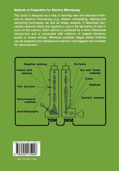
  • 1 An Introduction to Electron Microscopy (EM).- 1.1 Imaging Methods in Electron Microscopy.- 1.2 Preparation Procedures in TEM.- 1.3 Imaging Problems.- 1.4 Support Films.- 2 Methods for TEM.- 2.1 Fixation, Dehydration and Embedding.- 2.2 Ultramicrotomy.- 2.3 Macromolecular EM.- 2.4 Immunoelectron Microscopy (I EM).- 2.5 Autoradiography.- 2.6 Freeze (Fracturing) Etching.- 3 Methods for SEM.- 3.1 Conventional Methods of Preparation.- 3.2 Storage of Specimens.- 3.3 Demonstration of Surfaces via Replicas and Casts.- 3.4 Visualization of Internal Surfaces Through Sectioning and Dry-Fracturing (Dry-Cleaving).- 3.5 Element Analysis.- 4 Evaluation of Micrographs.- 4.1 Morphometry.- 4.2 Averaging and Image Reconstruction.- Appendix: Buffers in Electron Microscopy.