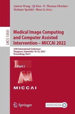
Medical Image Computing and Computer Assisted Intervention - MICCAI 2019 (eBook, PDF)
22nd International Conference, Shenzhen, China, October 13-17, 2019, Proceedings, Part VI
Redaktion: Shen, Dinggang; Khan, Ali; Yap, Pew-Thian; Zhou, Sean; Essert, Caroline; Staib, Lawrence H.; Peters, Terry M.; Liu, Tianming
Versandkostenfrei!
Sofort per Download lieferbar
72,95 €
inkl. MwSt.
Weitere Ausgaben:

PAYBACK Punkte
36 °P sammeln!
The six-volume set LNCS 11764, 11765, 11766, 11767, 11768, and 11769 constitutes the refereed proceedings of the 22nd International Conference on Medical Image Computing and Computer-Assisted Intervention, MICCAI 2019, held in Shenzhen, China, in October 2019.The 539 revised full papers presented were carefully reviewed and selected from 1730 submissions in a double-blind review process. The papers are organized in the following topical sections:Part I: optical imaging; endoscopy; microscopy.Part II: image segmentation; image registration; cardiovascular imaging; growth, development, atrophy a...
The six-volume set LNCS 11764, 11765, 11766, 11767, 11768, and 11769 constitutes the refereed proceedings of the 22nd International Conference on Medical Image Computing and Computer-Assisted Intervention, MICCAI 2019, held in Shenzhen, China, in October 2019.
The 539 revised full papers presented were carefully reviewed and selected from 1730 submissions in a double-blind review process. The papers are organized in the following topical sections:
Part I: optical imaging; endoscopy; microscopy.
Part II: image segmentation; image registration; cardiovascular imaging; growth, development, atrophy and progression.
Part III: neuroimage reconstruction and synthesis; neuroimage segmentation; diffusion weighted magnetic resonance imaging; functional neuroimaging (fMRI); miscellaneous neuroimaging.
Part IV: shape; prediction; detection and localization; machine learning; computer-aided diagnosis; image reconstruction and synthesis.
Part V: computer assisted interventions; MIC meets CAI.
Part VI: computed tomography; X-ray imaging.
Dieser Download kann aus rechtlichen Gründen nur mit Rechnungsadresse in A, B, BG, CY, CZ, D, DK, EW, E, FIN, F, GR, HR, H, IRL, I, LT, L, LR, M, NL, PL, P, R, S, SLO, SK ausgeliefert werden.


