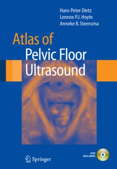
Ultrasound has replaced X-ray as the main imaging modality for the diagnosis of pelvic floor disorders in women. It now enables a cost-effective and non-invasive demonstration of bladder neck and pelvic organ mobility, vaginal, urethral and levator ani function and anatomy, and anorectal anatomy. Atlas of Pelvic Floor Ultrasound provides an introduction to pelvic floor imaging as well as a resource to be used during initial and more advanced practice.
Pelvic ? oor ultrasound is often described as a niche investigation within obstetrics and gynecology and even within gynecological ultrasound. After reading this book, I am convinced that it should be a mainstream investi- tion taught to all fellows and subspecialty trainees. Nothing should be of more importance to obstetricians and gynecologists than the protection of the pelvic ? oor of their patients and effective treatment when disorders arise. These disorders cause more prolonged and disruptive misery to patients than many of the conditions that clog up the waiting list in obst- rical and gynecological departments. This book is more than an "atlas"; it is an education in the anatomy and dynamics of the lower urinary tract and pelvic ? oor and the investigation of disorders that occur, such as incontinence and prolapse. Ultrasound, despite its preeminence as an investigative tool in obstetrics and gyne- logy, has been slow to achieve such status in urogynecology, principally because the transvaginal ultrasound probe which is the standard tool in gynecologic scanning distorts the pelvic ? oor anatomy. This makes int- pretation of prolapse impossible. It was the realization that perineal or translabial ultrasound provided equally good and artefact-free information on bladder dynamics and the integrity of the pelvic ? oor that a change in attitude occurred.
Pelvic ? oor ultrasound is often described as a niche investigation within obstetrics and gynecology and even within gynecological ultrasound. After reading this book, I am convinced that it should be a mainstream investi- tion taught to all fellows and subspecialty trainees. Nothing should be of more importance to obstetricians and gynecologists than the protection of the pelvic ? oor of their patients and effective treatment when disorders arise. These disorders cause more prolonged and disruptive misery to patients than many of the conditions that clog up the waiting list in obst- rical and gynecological departments. This book is more than an "atlas"; it is an education in the anatomy and dynamics of the lower urinary tract and pelvic ? oor and the investigation of disorders that occur, such as incontinence and prolapse. Ultrasound, despite its preeminence as an investigative tool in obstetrics and gyne- logy, has been slow to achieve such status in urogynecology, principally because the transvaginal ultrasound probe which is the standard tool in gynecologic scanning distorts the pelvic ? oor anatomy. This makes int- pretation of prolapse impossible. It was the realization that perineal or translabial ultrasound provided equally good and artefact-free information on bladder dynamics and the integrity of the pelvic ? oor that a change in attitude occurred.
From the reviews:
"This book takes a novice to the use of ultrasound in urogynaecology through an amazing journey exploring the use of this modality in assessing female patients with pelvic floor dysfunction. Its simplicity, rich practical detail and clear images will persuade the reader of the value of this investigation in day-to-day clinical practice. ... The book is an indispensable resource to all urogynaecologists who are keen to provide the best care for their patients." (Sharif I. M. F. Ismail, International Urogynaecology Journal, Vol. 20, 2009)
"This book takes a novice to the use of ultrasound in urogynaecology through an amazing journey exploring the use of this modality in assessing female patients with pelvic floor dysfunction. Its simplicity, rich practical detail and clear images will persuade the reader of the value of this investigation in day-to-day clinical practice. ... The book is an indispensable resource to all urogynaecologists who are keen to provide the best care for their patients." (Sharif I. M. F. Ismail, International Urogynaecology Journal, Vol. 20, 2009)

