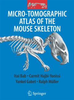
The Micro-Tomographic Atlas of the Mouse Skeleton provides a unique systematic description of all calcified components of the mouse. It includes about 200 high resolution, two and three dimensional m CT images of the exterior and interiors of all bones and joints. In addition, the spatial relationship of bones within complex skeletal units is also described. The images are accompanied by detailed explanatory text, thus highlighting special features and newly reported structures. The Atlas fulfils an emerging need for a comprehensive reference to assist both trained and in-training researchers.
From the reviews:
"The format of the book is clearly defined and well organized by the authors. ... serve as an important visual reference for researchers using mouse models in skeletal biology research. The Micro-Tomographic Atlas of the Mouse Skeleton is a great elementary reference to any collection and wonderfully illustrates the tremendous power of micro-CT technology in the imaging of the rodent skeleton." (Steven M. Tommasini and Christopher Price, Journal of Mammalian Evolution, Vol. 16, 2009)
"The format of the book is clearly defined and well organized by the authors. ... serve as an important visual reference for researchers using mouse models in skeletal biology research. The Micro-Tomographic Atlas of the Mouse Skeleton is a great elementary reference to any collection and wonderfully illustrates the tremendous power of micro-CT technology in the imaging of the rodent skeleton." (Steven M. Tommasini and Christopher Price, Journal of Mammalian Evolution, Vol. 16, 2009)


